

X-Ray & Fluoroscopy
United Regional offers advanced diagnostic imaging through X-ray and fluoroscopy — a real-time imaging technique that uses continuous X-rays — ensuring timely and accurate diagnoses.
If your provider orders an X-ray, you can conveniently schedule your appointment through MyChart at a time that works best for you.
How can United Regional’s X-ray & fluoroscopy services help you?
At United Regional, we’re happy to provide the following services, so that you may begin your healing as quickly as possible:
X-ray
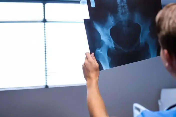
X-ray imaging uses invisible electromagnetic energy beams to produce detailed images of bones, organs, and internal tissues. At United Regional, X-rays are performed using external radiation that passes through the body and captures digital images on specially treated plates. These images appear in varying shades of black and white, depending on how different tissues absorb the radiation. X-rays are a foundational diagnostic tool, supporting timely and accurate clinical decisions.
Fluoroscopy
Fluoroscopy is a dynamic imaging technique that captures real-time movement of internal structures using continuous X-ray beams. Unlike traditional X-rays that produce still images, fluoroscopy functions like a video, allowing our providers to observe your organs and systems in motion. This method is particularly useful for evaluating cardiovascular, reproductive, and digestive systems. At United Regional, fluoroscopy is often used during barium procedures to see how contrast material moves through your body in real time. This real-time view helps your care team make more accurate diagnoses and plan the right treatment
Upper GI Series/Barium Swallow
An upper GI (gastrointestinal) series, sometimes called a barium swallow, uses real-time X-Ray imaging called fluoroscopy to examine your esophagus, stomach, and the first part of your small intestine (duodenum).
Your provider may order an upper GI series to evaluate how your upper digestive tract looks and functions. The exam can help identify reflux, ulcers, narrowing (strictures), hiatal hernia, and other abnormalities. It may also be used to evaluate symptoms such as abdominal pain, nausea or vomiting, difficulty swallowing, unexplained weight loss, or gastrointestinal bleeding.
What to Expect in an Upper GI Series
- You will drink a barium contrast liquid that coats your upper digestive tract and makes it easier to see on X-Ray.
- For some exams, you may also swallow fizzy crystals that create gas to help improve the images.
- A radiologist will use fluoroscopy to watch the contrast move through your esophagus, stomach, and duodenum and will take X-Ray images.
- You may be asked to change positions or briefly hold your breath so different areas can be viewed.
Barium Enema
A barium enema, also called a lower GI series, uses X-Ray and fluoroscopy to examine the large intestine (colon) and rectum. Barium contrast helps make the colon more visible on X-Ray.
A barium enema may be used to evaluate abdominal pain, constipation, chronic diarrhea, changes in bowel habits, rectal bleeding, or unexplained weight loss. It can also help identify certain abnormalities of the colon.
What to Expect During a Barium Enema
- You will lie on an X-Ray table.
- A barium contrast will be introduced into the colon via a small tube gently inserted into the rectum.
- Air may also be introduced to expand the colon and provide additional detail.
- A radiologist will use fluoroscopy and X-Rays to examine your colon. You may be asked to change positions so different areas can be viewed.
- You may experience pressure, cramping, or the urge to have a bowel movement during the exam.
Swallowing Function
A swallowing function exam, also called a Modified Barium Swallow (MBS), uses real-time X-Ray imaging called fluoroscopy to evaluate how safely and effectively you swallow.
This exam is commonly used for people who have difficulty swallowing, also known as dysphagia. It evaluates how food and liquids move through the mouth and throat and can help determine whether food or liquid is entering the airway, known as aspiration.
What to Expect During a Swallowing Function
- You will be given liquids and foods of different consistencies that are mixed or coated with barium so they can be seen on X-Ray.
- Fluoroscopy will record your swallowing in real time as you eat and drink.
- A speech-language pathologist will perform the exam and evaluate your swallowing function.
- Different positions or swallowing techniques may be tested to determine whether they help you swallow more safely.
Lumbar Puncture
A lumbar puncture, also called a spinal tap, is a procedure that may use fluoroscopy to guide a thin needle into the lower back to access cerebrospinal fluid (CSF), the fluid surrounding the spinal cord.
A lumbar puncture may be performed to collect a sample of CSF for laboratory testing or to measure CSF pressure. Testing the fluid can help diagnose infections and certain inflammatory or neurological conditions
What to Expect During a Lumbar Puncture
- You will be positioned on your stomach on an X-Ray table.
- Your lower back will be cleaned, and local anesthetic will be used to numb the area.
- Using fluoroscopy for guidance, a provider will carefully insert a thin needle into the lower spinal canal.
- A sample of spinal fluid will be collected and sent to the laboratory. Fluid pressure may also be measured if ordered.
Myelogram
A myelogram is a specialized imaging exam that uses fluoroscopy and contrast media to create detailed images of the spinal canal, spinal cord, and nerve roots.
A myelogram can help identify problems affecting the spinal cord or nerves, including spinal stenosis, herniated discs, and compression of the nerves or spinal cord.
What to Expect from a Myelogram
- You will be positioned on your stomach on an X-Ray table. Your lower back will be cleaned and numbed with local anesthetic.
- Using fluoroscopy for guidance, a needle will be placed into the spinal canal. Contrast media will then be injected into the fluid-filled space surrounding the spinal cord and nerves.
- A radiologist will use fluoroscopy to monitor the contrast and take X-Ray images of your spine.
- A CT scan will be performed afterward to provide more detailed images.
Arthrogram
An arthrogram is an imaging procedure used to examine the inside of a joint, such as the shoulder, hip, wrist, elbow, or knee. Contrast media is injected directly into the joint using X-Ray guidance to make the joint structures easier to see.
An arthrogram can help evaluate joint pain; injury; and instability or abnormalities involving cartilage, ligaments ,or the joint capsule. The procedure is commonly performed before an MRI or CT scan to provide additional detail of the joint.
What to Expect from an Arthrogram
- The skin over the joint will be cleaned and numbed with local anesthetic.
- Using fluoroscopy for guidance, a small needle will be carefully placed into the joint.
- Contrast media will be injected into the joint.
- X-Ray images may be taken. Depending on the exam ordered, you may then have an MRI or CT scan.
- Your joint may feel full or mildly uncomfortable after the contrast is injected.
Steroid Injection
A fluoroscopy-guided steroid injection uses real-time X-Ray imaging to help a radiologist accurately place medication into a joint or another targeted area. A small amount of contrast may also be used to confirm the needle's position.
A steroid injection may be used to reduce inflammation and relieve pain from conditions such as arthritis. Fluoroscopy helps the radiologist accurately place the medication in the targeted area.
What to Expect During a Fluoroscopy-Guided Steroid Injection
- You will be positioned on an X-Ray table, and the area will be cleaned.
- The skin may be numbed with a local anesthetic.
- Using fluoroscopy, the radiologist will guide a small needle into the appropriate location.
- A small amount of contrast may be injected to confirm the needle’s placement.
- Steroid medication and numbing medication are then injected.

Related Services and Technology

CarePlus
Convenient care for minor illnesses and injuries. Same-day care through CarePlus is available—either by walk-in or scheduled appointment—at the Barnett Road Medical Building in Wichita Falls.
Imaging
Accredited imaging services at United Regional ensure precision, safety, and expert care using advanced technologies.

Primary Care
Primary care is whole-person care that supports your physical, mental, and emotional health through prevention, diagnosis, treatment, and long-term wellness. Take your first step to long-term health.